Pathophysiology of Paranasal Aspergillus Colonization
The human paranasal sinuses are naturally sterile cavities lined with pseudostratified ciliated columnar epithelium. When individuals occupy indoor spaces with chronic Aspergillus colonization across organic substrates (such as damp gypsum drywall, Kraft paper insulation, and AC evaporator coil drip pans), microscopic conidia.
The human paranasal sinuses are naturally sterile cavities lined with pseudostratified ciliated columnar epithelium.
When individuals occupy indoor spaces with chronic colonization across organic substrates (such as damp gypsum drywall, Kraft paper insulation, and AC evaporator coil drip pans), microscopic conidia (measuring 2.0 to 3.5 microns in aerodynamic diameter) penetrate the narrow sinus ostia.
In patients with anatomical predispositions (such as deviated nasal septa, mucosal edema, or past dental surgery involving the maxillary sinus floor), the trapped spores germinate.
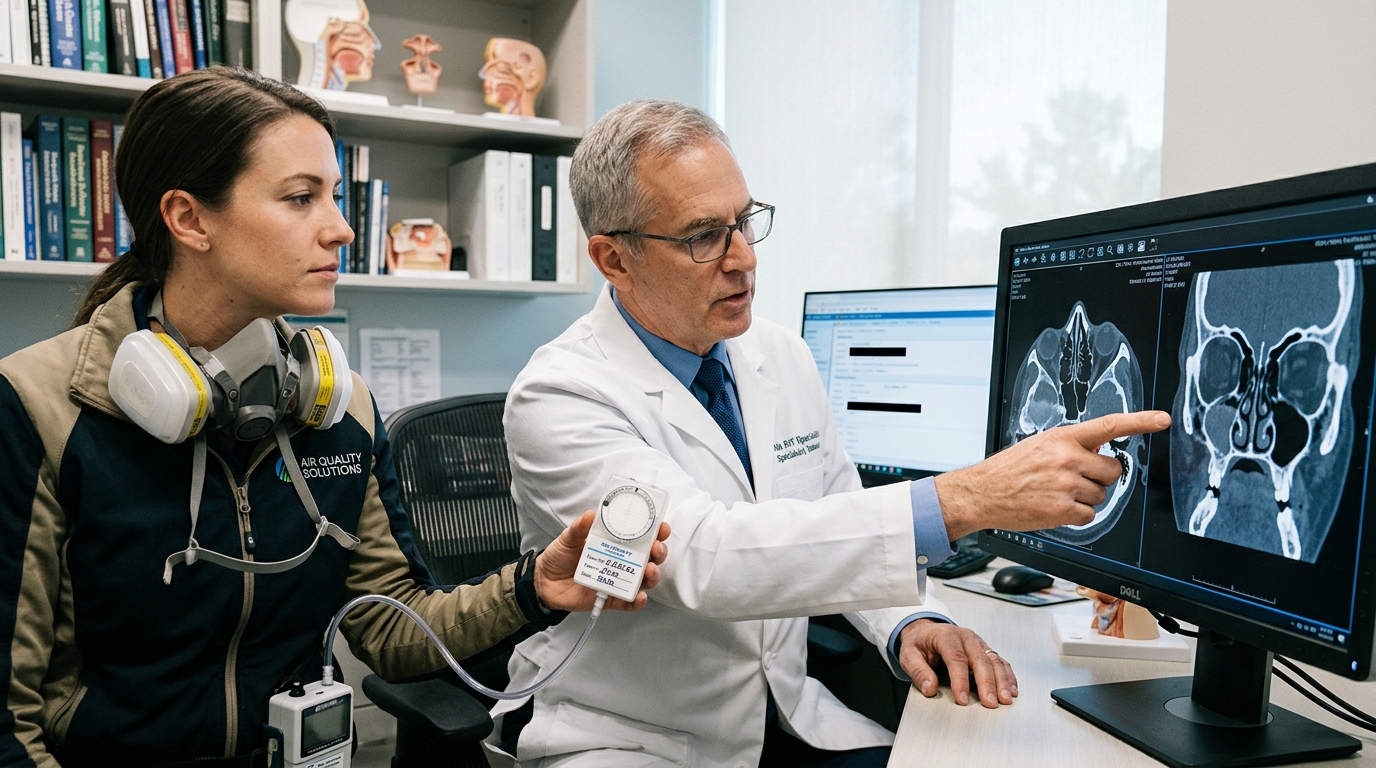
As fungal hyphae branch at characteristic 45-degree angles, they form a dense, tangled concretion resembling clay or wet papier-mâché, incorporating heavy calcium phosphate salts that appear as hyperdense, metallic calcifications on non-contrast maxillofacial CT scans.
Diagnostic Differentiation: Bacterial Sinusitis vs. Fungal Mycetoma
Otolaryngologists (ENT specialists) and environmental physicians differentiate fungal sinus balls from chronic rhinosinusitis using high-resolution coronal CT imaging and endoscopic visualization. Non-contrast CT scans reveal complete opacification of the affected maxillary antrum with central punctate calcifications and reactive osteosclerotic thickening of the.
Otolaryngologists (ENT specialists) and environmental physicians differentiate fungal sinus balls from chronic rhinosinusitis using high-resolution coronal CT imaging and endoscopic visualization.
Non-contrast CT scans reveal complete opacification of the affected maxillary antrum with central punctate calcifications and reactive osteosclerotic thickening of the surrounding sinus bony walls.
Unlike invasive fungal sinusitis seen in severely immunocompromised patients, fungal sinus balls do not exhibit tissue erosion into the orbit or cranial cavity; however, chronic fungal enzyme release causes localized mucosal inflammation and persistent systemic mycotoxin exposure.
Surgical Debridement & Medical Protocols
Definitive medical eradication of a fungal sinus ball requires Functional Endoscopic Sinus Surgery (FESS) performed under general anesthesia. The otolaryngologist performs a wide middle meatal antrostomy to expose the maxillary sinus cavity, thoroughly aspirating the brittle fungal debris, irrigating the cavity with.
Definitive medical eradication of a fungal sinus ball requires Functional Endoscopic Sinus Surgery (FESS) performed under general anesthesia.
The otolaryngologist performs a wide middle meatal antrostomy to expose the maxillary sinus cavity, thoroughly aspirating the brittle fungal debris, irrigating the cavity with pressurized saline, and sending biopsy specimens for Gomori methenamine silver (GMS) histological staining.
Because systemic oral antifungals (such as itraconazole or voriconazole) exhibit poor tissue penetration into non-vascularized fungal masses, surgical evacuation is mandatory.
Home Environmental Source Removal & HVAC Decontamination Standards
Post-surgical recovery fails completely if the patient returns to a mold-contaminated residence. Eradicating indoor fungal reservoirs requires certified environmental remediation: (1) establishing negative-pressure containment (-5 Pascals) using commercial HEPA 500 air scrubbers, (2) cutting out mold-damaged drywall and carpet underlayments, (3) decontaminating.
Post-surgical recovery fails completely if the patient returns to a mold-contaminated residence.
Eradicating indoor fungal reservoirs requires certified environmental remediation: (1) establishing negative-pressure containment (-5 Pascals) using commercial HEPA 500 air scrubbers, (2) cutting out mold-damaged drywall and carpet underlayments, (3) decontaminating central HVAC air handlers and ductwork under NADCA Standard ACR 2021 protocols, and (4) conducting third-party Post-Remediation Verification (PRV) spore trap air sampling at 15.0 liters per minute to guarantee Condition 1 normal fungal ecology.